

A Transportation System Consists Of:
| Circulatory system | |
|---|---|
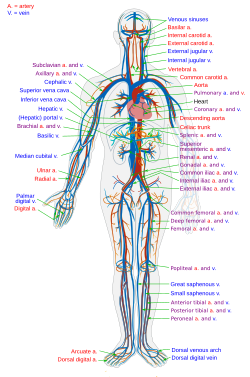 The human circulatory system (simplified). Ruby indicates oxygenated blood carried in arteries. Blue indicates deoxygenated claret carried in veins. Capillaries bring together the arteries and veins. | |
| Identifiers | |
| MeSH | D002319 |
| TA98 | A12.0.00.000 |
| TA2 | 3891 |
| FMA | 7161 |
| Anatomical terminology [edit on Wikidata] | |
The claret circulatory arrangement is a system of organs that includes the center, blood vessels, and claret which is circulated throughout the entire body of a human or other vertebrate.[1] [2] It includes the cardiovascular arrangement, or vascular system, that consists of the middle and claret vessels (from Greek kardia significant heart, and from Latin vascula meaning vessels). The circulatory system has two divisions, a systemic circulation or circuit, and a pulmonary circulation or circuit.[three] Some sources use the terms cardiovascular organisation and vascular system interchangeably with the circulatory arrangement.[4]
The network of blood vessels are the swell vessels of the heart including big elastic arteries, and big veins; other arteries, smaller arterioles, capillaries that join with venules (minor veins), and other veins. The circulatory organization is airtight in vertebrates, which means that the blood never leaves the network of claret vessels. Some invertebrates such equally arthropods have an open circulatory organisation. Diploblasts such as sponges, and comb jellies lack a circulatory system.
Blood is a fluid consisting of plasma, carmine blood cells, white blood cells, and platelets that is circulated around the torso carrying oxygen and nutrients to the tissues, and waste materials away. Circulated nutrients include proteins and minerals, other components transported are gases such equally oxygen, and carbon dioxide, hormones, and hemoglobin; providing nourishment, help in the immune system to fight diseases, and in maintaining homeostasis by stabilizing temperature and natural pH.
In vertebrates, complementary to the circulatory system is the lymphatic system. This system carries excess plasma filtered from the capillaries as interstitial fluid between cells, away from the torso tissues in an accessory route to return the excess fluid back to the claret apportionment every bit lymph.[five] The passage of lymph takes much longer than that of claret.[half dozen] The lymphatic system is a subsystem that is essential for the functioning of the claret circulatory organisation; without it the blood would become depleted of fluid. The lymphatic system works together with the allowed system.[7] Dissimilar the closed circulatory system, the lymphatic system is an open organisation. Some sources draw information technology as a secondary circulatory arrangement.
The circulatory system tin can be affected by many cardiovascular diseases. Cardiologists are medical professionals which specialise in the heart, and cardiothoracic surgeons specialise in operating on the centre and its surrounding areas. Vascular surgeons focus on disorders of the blood vessels, and lymphatic vessels.
Structure

Blood catamenia in the pulmonary and systemic circulations showing capillary networks in the torso sections
The circulatory organization includes the middle, blood vessels, and blood.[two] The cardiovascular arrangement in all vertebrates, consists of the heart and blood vessels. The circulatory system is farther divided into 2 major circuits – a pulmonary circulation, and a systemic circulation.[viii] [1] [iii] The pulmonary circulation is a circuit loop from the right heart taking deoxygenated blood to the lungs where information technology is oxygenated and returned to the left heart. The systemic apportionment is a excursion loop that delivers oxygenated blood from the left centre to the rest of the body, and returns deoxygenated blood dorsum to the right heart via large veins known every bit the venae cavae. The systemic circulation can likewise exist defined as 2 parts – a macrocirculation and a microcirculation. An average adult contains v to six quarts (roughly 4.7 to 5.7 liters) of blood, accounting for approximately 7% of their total body weight.[9] Blood consists of plasma, red blood cells, white claret cells, and platelets. The digestive organization also works with the circulatory system to provide the nutrients the arrangement needs to keep the centre pumping.[10]
Further circulatory routes are associated, such as the coronary apportionment to the middle itself, the cerebral circulation to the brain, renal circulation to the kidneys, and bronchial circulation to the bronchi in the lungs.
The homo circulatory organisation is closed, meaning that the blood is contained inside the vascular network.[xi] Nutrients travel through tiny blood vessels of the microcirculation to reach organs.[xi] The lymphatic system is an essential subsystem of the circulatory system consisting of a network of lymphatic vessels, lymph nodes, organs, tissues and circulating lymph. This subsystem is an open organisation.[12] A major office is to carry the lymph, draining and returning interstitial fluid into the lymphatic ducts back to the heart for return to the circulatory system. Another major role is working together with the allowed system to provide defense against pathogens.[13]
Heart
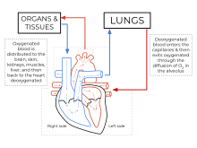
Diagram of the human heart showing blood oxygenation to the pulmonary and systemic circulation
The heart pumps blood to all parts of the body providing nutrients and oxygen to every cell, and removing waste products. The left heart pumps oxygenated blood returned from the lungs to the rest of the body in the systemic circulation. The right heart pumps deoxygenated blood to the lungs in the pulmonary apportionment. In the human heart there is one atrium and one ventricle for each circulation, and with both a systemic and a pulmonary circulation in that location are four chambers in total: left atrium, left ventricle, right atrium and right ventricle. The right atrium is the upper bedchamber of the right side of the heart. The claret that is returned to the correct atrium is deoxygenated (poor in oxygen) and passed into the right ventricle to exist pumped through the pulmonary artery to the lungs for re-oxygenation and removal of carbon dioxide. The left atrium receives newly oxygenated claret from the lungs likewise as the pulmonary vein which is passed into the potent left ventricle to be pumped through the aorta to the different organs of the body.
Pulmonary circulation

The pulmonary circulation is the portion of the cardiovascular arrangement in which oxygen-depleted blood is pumped abroad from the center, via the pulmonary artery, to the lungs and returned, oxygenated, to the heart via the pulmonary vein.
Oxygen-deprived claret from the superior and junior vena cava enters the correct atrium of the heart and flows through the tricuspid valve (right atrioventricular valve) into the right ventricle, from which it is then pumped through the pulmonary semilunar valve into the pulmonary artery to the lungs. Gas exchange occurs in the lungs, whereby COtwo is released from the blood, and oxygen is absorbed. The pulmonary vein returns the now oxygen-rich blood to the left atrium.[10]
A carve up system known equally the bronchial circulation supplies blood to the tissue of the larger airways of the lung.
Systemic circulation


Diagram of capillary network joining the arterial system with the venous system.
The systemic circulation is a excursion loop that delivers oxygenated blood from the left heart to the remainder of the trunk through the aorta. Deoxygenated claret is returned in the systemic apportionment to the correct middle via ii large veins, the junior vena cava and the superior vena cava, where information technology is pumped from the right atrium into the pulmonary circulation for oxygenation. The systemic apportionment can also be defined as having two parts – a macrocirculation and a microcirculation.[10]
Blood vessels
The blood vessels of the circulatory organisation are the arteries, veins, and capillaries. The large arteries and veins that accept blood to, and abroad from the middle are known as the groovy vessels.[xiv]
Arteries
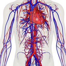
Delineation of the heart, major veins and arteries constructed from body scans
Oxygenated claret enters the systemic circulation when leaving the left ventricle, through the aortic semilunar valve.[fifteen] The kickoff part of the systemic apportionment is the aorta, a massive and thick-walled artery. The aorta arches and gives branches supplying the upper part of the trunk after passing through the aortic opening of the diaphragm at the level of thoracic ten vertebra, information technology enters the abdomen.[16] Subsequently, it descends down and supplies branches to abdomen, pelvis, perineum and the lower limbs.[17]
The walls of the aorta are elastic. This elasticity helps to maintain the claret pressure throughout the torso.[18] When the aorta receives almost five litres of blood from the center, it recoils and is responsible for pulsating blood pressure. Equally the aorta branches into smaller arteries, their elasticity goes on decreasing and their compliance goes on increasing.[18]
Capillaries
Arteries branch into modest passages called arterioles and then into the capillaries.[19] The capillaries merge to bring blood into the venous organization.[20]
Veins
Capillaries merge into venules, which merge into veins.[21] The venous organisation feeds into the two major veins: the superior vena cava – which mainly drains tissues above the eye – and the inferior vena cava – which mainly drains tissues below the heart. These two large veins empty into the right atrium of the centre.[22]
Portal veins
The general dominion is that arteries from the heart branch out into capillaries, which collect into veins leading back to the centre. Portal veins are a slight exception to this. In humans the only pregnant example is the hepatic portal vein which combines from capillaries around the alimentary canal where the claret absorbs the various products of digestion; rather than leading directly back to the heart, the hepatic portal vein branches into a second capillary organization in the liver.
Coronary circulation
The heart itself is supplied with oxygen and nutrients through a small "loop" of the systemic apportionment and derives very lilliputian from the claret contained within the 4 chambers. The coronary apportionment arrangement provides a blood supply to the heart muscle itself. The coronary circulation begins near the origin of the aorta past 2 coronary arteries: the right coronary artery and the left coronary artery. After nourishing the heart muscle, blood returns through the coronary veins into the coronary sinus and from this i into the correct atrium. Back menses of blood through its opening during atrial systole is prevented by Thebesian valve. The smallest cardiac veins drain directly into the heart chambers.[x]
Cognitive circulation
The brain has a dual claret supply, an anterior and a posterior circulation from arteries at its front and dorsum. The inductive circulation arises from the internal carotid arteries to supply the front end of the encephalon. The posterior circulation arises from the vertebral arteries, to supply the dorsum of the brain and brainstem. The circulation from the front end and the dorsum bring together (anastomise) at the circumvolve of Willis.
Renal circulation
The renal circulation is the blood supply to the kidneys, contains many specialized blood vessels and receives around xx% of the cardiac output. It branches from the intestinal aorta and returns blood to the ascending vena cava.
Development
The evolution of the circulatory organization starts with vasculogenesis in the embryo. The human arterial and venous systems develop from different areas in the embryo. The arterial arrangement develops mainly from the aortic arches, six pairs of arches that develop on the upper role of the embryo. The venous system arises from three bilateral veins during weeks four – viii of embryogenesis. Fetal circulation begins within the 8th calendar week of evolution. Fetal apportionment does not include the lungs, which are bypassed via the truncus arteriosus. Earlier birth the fetus obtains oxygen (and nutrients) from the mother through the placenta and the umbilical cord.[23]
Arteries
Animation of a typical man reddish blood cell cycle in the circulatory arrangement. This animation occurs at a faster rate (~twenty seconds of the boilerplate 60-2d cycle) and shows the red claret cell deforming as it enters capillaries, as well equally the confined changing color every bit the prison cell alternates in states of oxygenation forth the circulatory organization.
The human arterial system originates from the aortic arches and from the dorsal aortae starting from week 4 of embryonic life. The first and second aortic arches regress and form only the maxillary arteries and stapedial arteries respectively. The arterial arrangement itself arises from aortic arches 3, 4 and 6 (aortic curvation v completely regresses).
The dorsal aortae, present on the dorsal side of the embryo, are initially nowadays on both sides of the embryo. They subsequently fuse to form the basis for the aorta itself. Approximately thirty smaller arteries branch from this at the back and sides. These branches form the intercostal arteries, arteries of the arms and legs, lumbar arteries and the lateral sacral arteries. Branches to the sides of the aorta will form the definitive renal, suprarenal and gonadal arteries. Finally, branches at the front of the aorta consist of the vitelline arteries and umbilical arteries. The vitelline arteries course the celiac, superior and junior mesenteric arteries of the gastrointestinal tract. Later on nascency, the umbilical arteries will form the internal iliac arteries.
Veins
The human venous arrangement develops mainly from the vitelline veins, the umbilical veins and the cardinal veins, all of which empty into the sinus venosus.
Part
Nearly 98.5% of the oxygen in a sample of arterial claret in a salubrious human being, breathing air at body of water-level force per unit area, is chemically combined with hemoglobin molecules. Virtually ane.5% is physically dissolved in the other claret liquids and not connected to hemoglobin. The hemoglobin molecule is the principal transporter of oxygen in vertebrates.
Clinical significance
Many diseases affect the circulatory arrangement. These include a number of cardiovascular diseases, affecting the heart and blood vessels; hematologic diseases that affect the claret, such as anemia, and lymphatic diseases affecting the lymphatic system. Cardiologists are medical professionals which specialise in the heart, and cardiothoracic surgeons specialise in operating on the heart and its surrounding areas. Vascular surgeons focus on the claret vessels.
Cardiovascular disease
Diseases affecting the cardiovascular organization are called cardiovascular disease.
Many of these diseases are called "lifestyle diseases" because they develop over time and are related to a person'south exercise habits, diet, whether they fume, and other lifestyle choices a person makes. Atherosclerosis is the precursor to many of these diseases. It is where small atheromatous plaques build upward in the walls of medium and large arteries. This may eventually grow or rupture to occlude the arteries. It is likewise a take a chance cistron for acute coronary syndromes, which are diseases that are characterised by a sudden arrears of oxygenated blood to the heart tissue. Atherosclerosis is besides associated with bug such every bit aneurysm germination or splitting ("autopsy") of arteries.
Another major cardiovascular disease involves the creation of a jell, called a "thrombus". These can originate in veins or arteries. Deep venous thrombosis, which mostly occurs in the legs, is one cause of clots in the veins of the legs, particularly when a person has been stationary for a long fourth dimension. These clots may embolise, meaning travel to some other location in the trunk. The results of this may include pulmonary embolus, transient ischaemic attacks, or stroke.
Cardiovascular diseases may also be congenital in nature, such equally centre defects or persistent fetal circulation, where the circulatory changes that are supposed to happen afterward birth do not. Not all congenital changes to the circulatory system are associated with diseases, a large number are anatomical variations.
Investigations
The function and health of the circulatory system and its parts are measured in a variety of manual and automated means. These include unproblematic methods such as those that are role of the cardiovascular examination, including the taking of a person's pulse as an indicator of a person's center rate, the taking of blood force per unit area through a sphygmomanometer or the utilize of a stethoscope to listen to the heart for murmurs which may bespeak problems with the middle's valves. An electrocardiogram can also be used to evaluate the mode in which electricity is conducted through the heart.
Other more invasive means tin can also be used. A cannula or catheter inserted into an avenue may be used to mensurate pulse pressure or pulmonary wedge pressures. Angiography, which involves injecting a dye into an artery to visualise an arterial tree, can be used in the eye (coronary angiography) or brain. At the aforementioned time every bit the arteries are visualised, blockages or narrowings may be stock-still through the insertion of stents, and active bleeds may be managed past the insertion of coils. An MRI may be used to epitome arteries, called an MRI angiogram. For evaluation of the blood supply to the lungs a CT pulmonary angiogram may exist used. Vascular ultrasonography may exist used to investigate vascular diseases affecting the venous arrangement and the arterial system including the diagnosis of stenosis, thrombosis or venous insufficiency. An intravascular ultrasound using a catheter is also an option.
Surgery
| | This section needs expansion. You can help past adding to it. (March 2015) |
There are a number of surgical procedures performed on the circulatory arrangement:
- Coronary artery bypass surgery
- Coronary stent used in angioplasty
- Vascular surgery
- Vein stripping
- Cosmetic procedures
Cardiovascular procedures are more likely to be performed in an inpatient setting than in an ambulatory intendance setting; in the U.s.a., only 28% of cardiovascular surgeries were performed in the ambulatory intendance setting.[24]
Other animals
The open circulatory system of the grasshopper – made upward of a heart, vessels and hemolymph. The hemolymph is pumped through the centre, into the aorta, dispersed into the head and throughout the hemocoel, and so back through the ostia in the heart and the process repeated.
While humans, also every bit other vertebrates, have a closed blood circulatory organization (significant that the claret never leaves the network of arteries, veins and capillaries), some invertebrate groups have an open circulatory system containing a heart but limited blood vessels. The most archaic, diploblastic animate being phyla lack circulatory systems.
An additional transport system, the lymphatic system, which is only plant in animals with a airtight blood apportionment, is an open arrangement providing an accompaniment road for excess interstitial fluid to be returned to the blood.[5]
The blood vascular organization offset appeared probably in an ancestor of the triploblasts over 600 million years ago, overcoming the time-altitude constraints of diffusion, while endothelium evolved in an bequeathed vertebrate some 540–510 million years ago.[25]
Open circulatory arrangement
In arthropods, the open up circulatory organisation is a system in which a fluid in a cavity called the hemocoel bathes the organs directly with oxygen and nutrients, with there existence no distinction betwixt claret and interstitial fluid; this combined fluid is called hemolymph or haemolymph.[26] Muscular movements past the beast during locomotion can facilitate hemolymph move, only diverting menstruation from i area to another is limited. When the center relaxes, claret is drawn back toward the centre through open up-ended pores (ostia).
Hemolymph fills all of the interior hemocoel of the torso and surrounds all cells. Hemolymph is composed of h2o, inorganic salts (mostly sodium, chloride, potassium, magnesium, and calcium), and organic compounds (mostly carbohydrates, proteins, and lipids). The primary oxygen transporter molecule is hemocyanin.
There are free-floating cells, the hemocytes, within the hemolymph. They play a role in the arthropod immune system.

Closed circulatory organisation

Ii-chambered heart of a fish
The circulatory systems of all vertebrates, as well as of annelids (for case, earthworms) and cephalopods (squids, octopuses and relatives) always go along their circulating blood enclosed within heart chambers or claret vessels and are classified every bit closed, simply as in humans. All the same, the systems of fish, amphibians, reptiles, and birds evidence various stages of the evolution of the circulatory arrangement.[27] Closed systems let blood to be directed to the organs that require it.
In fish, the system has merely one circuit, with the claret being pumped through the capillaries of the gills and on to the capillaries of the body tissues. This is known as single cycle circulation. The heart of fish is, therefore, but a single pump (consisting of two chambers).
In amphibians and most reptiles, a double circulatory arrangement is used, just the heart is not e'er completely separated into two pumps. Amphibians have a three-chambered heart.
In reptiles, the ventricular septum of the heart is incomplete and the pulmonary avenue is equipped with a sphincter muscle. This allows a 2nd possible road of blood catamenia. Instead of blood flowing through the pulmonary avenue to the lungs, the sphincter may exist contracted to divert this blood menses through the incomplete ventricular septum into the left ventricle and out through the aorta. This means the claret flows from the capillaries to the heart and back to the capillaries instead of to the lungs. This process is useful to ectothermic (cold-blooded) animals in the regulation of their body temperature.
Mammals, birds and crocodilians testify complete separation of the heart into 2 pumps, for a total of four heart chambers; it is idea that the four-chambered heart of birds and crocodilians evolved independently from that of mammals.[28] Double circulatory systems allow blood to exist repressurized later on returning from the lungs, speeding up delivery of oxygen to tissues.
No circulatory system
Circulatory systems are absent in some animals, including flatworms. Their body cavity has no lining or enclosed fluid. Instead, a muscular pharynx leads to an extensively branched digestive arrangement that facilitates direct diffusion of nutrients to all cells. The flatworm'south dorso-ventrally flattened trunk shape as well restricts the distance of any jail cell from the digestive organisation or the exterior of the organism. Oxygen can diffuse from the surrounding h2o into the cells, and carbon dioxide can diffuse out. Consequently, every cell is able to obtain nutrients, water and oxygen without the need of a send system.
Some animals, such every bit jellyfish, have more than all-encompassing branching from their gastrovascular cavity (which functions as both a place of digestion and a form of circulation), this branching allows for bodily fluids to reach the outer layers, since the digestion begins in the inner layers.
History

Human being anatomical nautical chart of claret vessels, with heart, lungs, liver and kidneys included. Other organs are numbered and bundled around information technology. Before cutting out the figures on this page, Vesalius suggests that readers glue the page onto parchment and gives instructions on how to assemble the pieces and paste the multilayered figure onto a base "musculus man" illustration. "Paradigm", fol.14a. HMD Collection, WZ 240 V575dhZ 1543.
The earliest known writings on the circulatory system are found in the Ebers Papyrus (16th century BCE), an ancient Egyptian medical papyrus containing over 700 prescriptions and remedies, both physical and spiritual. In the papyrus, it acknowledges the connection of the heart to the arteries. The Egyptians idea air came in through the mouth and into the lungs and heart. From the heart, the air travelled to every fellow member through the arteries. Although this concept of the circulatory system is only partially correct, information technology represents ane of the primeval accounts of scientific thought.
In the 6th century BCE, the knowledge of apportionment of vital fluids through the body was known to the Ayurvedic physician Sushruta in ancient India.[29] He also seems to have possessed knowledge of the arteries, described as 'channels' by Dwivedi & Dwivedi (2007).[29] The valves of the center were discovered by a physician of the Hippocratean schoolhouse effectually the fourth century BCE. However, their function was not properly understood then. Because claret pools in the veins later death, arteries expect empty. Ancient anatomists assumed they were filled with air and that they were for the transport of air.
The Greek doctor, Herophilus, distinguished veins from arteries but thought that the pulse was a property of arteries themselves. Greek anatomist Erasistratus observed that arteries that were cut during life drain. He ascribed the fact to the phenomenon that air escaping from an artery is replaced with blood that enters betwixt veins and arteries by very pocket-size vessels. Thus he apparently postulated capillaries but with reversed catamenia of blood.[ citation needed ]
In 2d-century AD Rome, the Greek dr. Galen knew that blood vessels carried blood and identified venous (night scarlet) and arterial (brighter and thinner) blood, each with distinct and separate functions. Growth and energy were derived from venous claret created in the liver from chyle, while arterial blood gave vitality past containing pneuma (air) and originated in the heart. Blood flowed from both creating organs to all parts of the trunk where it was consumed and there was no render of blood to the heart or liver. The middle did not pump blood effectually, the eye's motion sucked blood in during diastole and the blood moved by the pulsation of the arteries themselves.
Galen believed that the arterial blood was created by venous claret passing from the left ventricle to the right by passing through 'pores' in the interventricular septum, air passed from the lungs via the pulmonary artery to the left side of the heart. As the arterial blood was created 'sooty' vapors were created and passed to the lungs as well via the pulmonary avenue to be exhaled.
In 1025, The Canon of Medicine past the Persian physician, Avicenna, "erroneously accustomed the Greek notion regarding the beingness of a hole in the ventricular septum by which the blood traveled betwixt the ventricles." Despite this, Avicenna "correctly wrote on the cardiac cycles and valvular function", and "had a vision of blood circulation" in his Treatise on Pulse.[thirty] [ verification needed ] While besides refining Galen's erroneous theory of the pulse, Avicenna provided the first correct explanation of pulsation: "Every beat of the pulse comprises two movements and ii pauses. Thus, expansion : pause : contraction : pause. [...] The pulse is a movement in the eye and arteries ... which takes the form of alternating expansion and contraction."[31]
In 1242, the Arabian doc, Ibn al-Nafis described the procedure of pulmonary circulation in greater, more than accurate detail than his predecessors, though he believed, as they did, in the notion of vital spirit (pneuma), which he believed was formed in the left ventricle. Ibn al-Nafis stated in his Commentary on Beefcake in Avicenna'southward Catechism:
"...the blood from the correct chamber of the middle must make it at the left chamber simply there is no directly pathway between them. The thick septum of the heart is not perforated and does not have visible pores as some people thought or invisible pores as Galen thought. The blood from the right sleeping accommodation must period through the vena arteriosa (pulmonary artery) to the lungs, spread through its substances, be mingled there with air, pass through the arteria venosa (pulmonary vein) to achieve the left bedroom of the heart and there course the vital spirit..."
In add-on, Ibn al-Nafis had an insight into what would get a larger theory of the capillary circulation. He stated that "there must exist small communications or pores (manafidh in Arabic) between the pulmonary avenue and vein," a prediction that preceded the discovery of the capillary system by more than than 400 years.[32] Ibn al-Nafis' theory, however, was bars to blood transit in the lungs and did not extend to the entire trunk.
Michael Servetus was the outset European to depict the role of pulmonary circulation, although his achievement was non widely recognized at the fourth dimension, for a few reasons. He firstly described it in the "Manuscript of Paris"[33] [34] (near 1546), only this work was never published. And subsequently he published this description, just in a theological treatise, Christianismi Restitutio, not in a volume on medicine. Only three copies of the book survived just these remained hidden for decades, the balance were burned shortly afterward its publication in 1553 because of persecution of Servetus by religious authorities.
Better known discovery of pulmonary circulation was past Vesalius'southward successor at Padua, Realdo Colombo, in 1559.
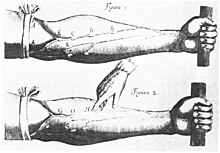
Finally, the English physician William Harvey, a pupil of Hieronymus Fabricius (who had earlier described the valves of the veins without recognizing their function), performed a sequence of experiments and published his Exercitatio Anatomica de Motu Cordis et Sanguinis in Animalibus in 1628, which "demonstrated that at that place had to be a direct connection between the venous and arterial systems throughout the torso, and not only the lungs. Most importantly, he argued that the crush of the heart produced a continuous circulation of blood through minute connections at the extremities of the body. This is a conceptual jump that was quite different from Ibn al-Nafis' refinement of the anatomy and bloodflow in the eye and lungs."[35] This work, with its essentially correct exposition, slowly convinced the medical world. Even so, Harvey was not able to identify the capillary system connecting arteries and veins; these were afterwards discovered by Marcello Malpighi in 1661.
In 1956, André Frédéric Cournand, Werner Forssmann and Dickinson Westward. Richards were awarded the Nobel Prize in Medicine "for their discoveries apropos heart catheterization and pathological changes in the circulatory arrangement."[36] In his Nobel lecture, Forssmann credits Harvey every bit birthing cardiology with the publication of his book in 1628.[37]
In the 1970s, Diana McSherry developed reckoner-based systems to create images of the circulatory system and center without the demand for surgery.[38]
Run into as well
- Cardiology – Branch of medicine dealing with the heart
- Cardiovascular drift
- Cardiac bike – Performance of the homo heart
- Vital heat
- Cardiac musculus – Muscular tissue of heart in vertebrates
- Major systems of the human body
- Amato Lusitano
- Vascular resistance – Force from blood vessels that affects claret flow
References
- ^ a b Hall, John E. (2011). Guyton and Hall textbook of medical physiology (Twelfth ed.). Philadelphia, Pa. p. iv. ISBN9781416045748.
- ^ a b Saladin, Kenneth S. (2011). Man anatomy (3rd ed.). New York: McGraw-Colina. p. 520. ISBN9780071222075.
- ^ a b Saladin, Kenneth Southward. (2011). Man beefcake (third ed.). New York: McGraw-Hill. p. 540. ISBN9780071222075.
- ^ How does the blood circulatory system work? - InformedHealth.org - NCBI Bookshelf. Institute for Quality and Efficiency in Health Intendance (IQWiG). 31 January 2019. Archived from the original on 29 January 2022.
- ^ a b Sherwood, Lauralee (2011). Human Physiology: From Cells to Systems. Cengage Learning. pp. 401–. ISBN978-1-133-10893-one. Archived from the original on 2020-07-29. Retrieved 2015-06-27 .
- ^ "The lymphatic arrangement and cancer | Cancer Research UK". 29 October 2014. Archived from the original on 30 Jan 2022. Retrieved January 30, 2022.
- ^ Saladin, Kenneth S. (2011). Human beefcake (3rd ed.). New York: McGraw-Colina. p. 610. ISBN9780071222075.
- ^ Cardiovascular+System at the US National Library of Medicine Medical Subject Headings (MeSH)
- ^ Pratt, Rebecca. "Cardiovascular System: Blood". AnatomyOne. Amirsys, Inc. Archived from the original on 2017-02-24.
- ^ a b c d Guyton, Arthur; Hall, John (2000). Guyton Textbook of Medical Physiology (10 ed.). ISBN978-0-7216-8677-6.
- ^ a b Lawton, Cassie M. (2019). The Human Circulatory Arrangement. Cavendish Foursquare Publishing. p. six. ISBN978-1-50-265720-6. Archived from the original on 2022-01-28. Retrieved 2022-01-28 .
- ^ Gartner, Leslie P.; Hiatt, James 50. (2010). Concise Histology Due east-Volume. Elsevier Health Sciences. p. 166. ISBN978-1-43-773579-6. Archived from the original on 2022-01-28. Retrieved 2022-01-28 .
- ^ Alberts, B.; Johnson, A.; Lewis, J.; Raff, M.; Roberts, Thou.; Walters, P. (2002). Molecular Biology of the Cell (fourth ed.). New York and London: Garland Science. ISBN978-0-8153-3218-3. Archived from the original on 2006-08-17. Retrieved 2017-08-thirty .
- ^ Standring, Susan (2016). Gray'south anatomy : the anatomical basis of clinical practice (Xl-offset ed.). [Philadelphia]. p. 1024. ISBN9780702052309.
- ^ Iaizzo, Paul A (2015). Handbook of Cardiac Beefcake, Physiology, and Devices. Springer. p. 93. ISBN978-3-31919464-6. Archived from the original on 2017-x-eleven. Retrieved 2022-01-28 .
- ^ Iaizzo, Paul A (2015). Handbook of Cardiac Anatomy, Physiology, and Devices. Springer. pp. v, 77. ISBN978-three-31919464-half-dozen. Archived from the original on 2017-10-11. Retrieved 2022-01-28 .
- ^ Iaizzo, Paul A (2015). Handbook of Cardiac Anatomy, Physiology, and Devices. Springer. pp. 5, 41–43. ISBN978-3-31919464-6. Archived from the original on 2017-10-11. Retrieved 2022-01-28 .
- ^ a b Vaz, Mario; Raj, Toni; Anura, Kurpad (2016). Guyton & Hall Textbook of Medical Physiology - Due east-Book: A South Asian Edition. Elsevier Health Sciences. p. 255. ISBN978-8-xiii-124665-8. Archived from the original on 2022-01-28. Retrieved 2022-01-28 .
- ^ National Institutes of Health. "What Are the Lungs?". nih.gov. Archived from the original on 2014-10-04.
- ^ State Academy of New York (February 3, 2014). "The Circulatory System". suny.edu. Archived from the original on February 3, 2014.
- ^ Mcconnell, Thomas H.; Hull, Kerry Fifty. (2020). Human Class, Human Office: Essentials of Anatomy & Physiology, Enhanced Edition. Jones & Bartlett Learning. p. 432. ISBN978-1-28-421805-three. Archived from the original on 2022-01-28. Retrieved 2022-01-28 .
- ^ Parkinson, Clayton Floyd; Huether, Sue E.; McCance, Kathryn 50. (2000). Agreement Pathophysiology. Mosby. p. 161. ISBN978-0-32-300792-iv.
- ^ Whitaker, Kent (2001). "Fetal Apportionment". Comprehensive Perinatal and Pediatric Respiratory Care. Delmar Thomson Learning. pp. 18–20. ISBN978-0-7668-1373-1. [ permanent dead link ]
- ^ Wier LM, Steiner CA, Owens PL (April 17, 2015). "Surgeries in Hospital-Owned Outpatient Facilities, 2012". HCUP Statistical Brief #188. Rockville, Physician: Bureau for Healthcare Research and Quality. Archived from the original on March 12, 2015.
- ^ Monahan-Earley, R.; Dvorak, A. M.; Aird, Westward. C. (2013). "Evolutionary origins of the blood vascular system and endothelium". Journal of Thrombosis and Haemostasis. 11: 46–66. doi:x.1111/jth.12253. PMC5378490. PMID 23809110.
- ^ Bailey, Regina. "Circulatory Organisation". biology.nigh.com. Archived from the original on 2016-eleven-29. Retrieved 2022-02-23 .
- ^ Simões-Costa, Marcos S.; Vasconcelos, Michelle; Sampaio, Allysson C.; Cravo, Roberta One thousand.; Linhares, Vania 50.; Hochgreb, Tatiana; Yan, Chao Y.I.; Davidson, Brad; Xavier-Neto, José (2005). "The evolutionary origin of cardiac chambers". Developmental Biology. 277 (1): 1–15. doi:10.1016/j.ydbio.2004.09.026. PMID 15572135.
- ^ "Crocodilian Hearts". National Centre for Science Education. October 24, 2008. Archived from the original on September 26, 2015. Retrieved Oct iii, 2015.
- ^ a b Dwivedi, Girish & Dwivedi, Shridhar (2007). "History of Medicine: Sushruta – the Clinician – Instructor par Excellence" Archived October 10, 2008, at the Wayback Machine, Indian J Chest Dis Allied Sci Vol. 49 pp. 243–244, National Informatics Center (Authorities of India).
- ^ Shoja, M.Thou.; Tubbs, R.S.; Loukas, M.; Khalili, M.; Alakbarli, F.; Cohen-Gadol, A.A. (2009). "Vasovagal syncope in the Canon of Avicenna: The beginning mention of carotid artery hypersensitivity". International Journal of Cardiology. 134 (3): 297–301. doi:ten.1016/j.ijcard.2009.02.035. PMID 19332359.
- ^ Hajar, Rachel (1999). "The Greco-Islamic Pulse". Middle Views. 1 (4): 136–140 [138]. Archived from the original on 2014-01-09.
- ^ West, J.B. (2008). "Ibn al-Nafis, the pulmonary circulation, and the Islamic Golden Age". Journal of Applied Physiology. 105 (vi): 1877–1880. doi:x.1152/japplphysiol.91171.2008. PMC2612469. PMID 18845773.
- ^ Gonzalez Etxeberria, Patxi (2011) Amor a la verdad, el – vida y obra de Miguel servet [The love for truth. Life and work of Michael Servetus]. Navarro y Navarro, Zaragoza, collaboration with the Government of Navarra, Department of Institutional Relations and Instruction of the Government of Navarra. ISBN 84-235-3266-half dozen pp. 215–228 & 62nd illustration (XLVII)
- ^ Michael Servetus Enquiry Archived 2012-xi-13 at the Wayback Auto Study with graphical proof on the Manuscript of Paris and many other manuscripts and new works by Servetus
- ^ Pormann, Peter Due east. and Smith, East. Roughshod (2007) Medieval Islamic medicine Georgetown University, Washington DC, p. 48, ISBN one-58901-161-9.
- ^ "The Nobel Prize in Physiology or Medicine 1956". Nobel Foundation. Archived from the original on 2007-09-29. Retrieved 2007-07-28 .
- ^ "The Function of Middle Catheterization and Angiocardiography in the Evolution of Mod Medicine". Archived from the original on 2017-10-09. Retrieved 2017-10-08 .
- ^ Wayne, Tiffany Chiliad. (2011). American women of scientific discipline since 1900 . Santa Barbara, Calif.: ABC-CLIO. pp. 677–678. ISBN978-1-59884-158-ix.
External links
- Circulatory Pathways in Anatomy and Physiology by OpenStax
- The Circulatory System
- Michael Servetus Research Study on the Manuscript of Paris by Servetus (1546 description of the Pulmonary Circulation)

A Transportation System Consists Of:,
Source: https://en.wikipedia.org/wiki/Circulatory_system
Posted by: richardsonnounkilthe.blogspot.com
0 Response to "A Transportation System Consists Of:"
Post a Comment